| Info
Sheets |
| | | | | | | | | | | | | | | | | | | | | | | | |
 | Out-
side |
| | | | |
|
| | | | |
Result : Searchterm 'MRI of the Brain' found in 0 term [ ] and 0 definition [ ] and 0 definition [ ], (+ 20 Boolean[ ], (+ 20 Boolean[ ] results ] results
| | previous 6 - 10 (of 20) nextResult Pages : [1 2 3 4] |  | |  | Searchterm 'MRI of the Brain' was also found in the following services: | | | | |
| |  |
| |
|
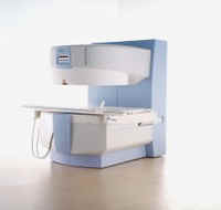
Device Information and Specification
CLINICAL APPLICATION
Whole body
GRE, IR, FIR, STIR, TrueIR/FISP, FSE, MT, SS-FSE, MT-SE, MTC, MSE, GMR, fat/water sat./exc.
IMAGING MODES
Single, multislice, volume study, multi angle
512 x 512 full screen display
POWER REQUIREMENTS
380/400/420/440/480 V
| | | | | | | | | | |
| | | | | |
| |
|
Contrast enhanced MRI is a commonly used procedure in magnetic resonance imaging. The need to more accurately characterize different types of lesions and to detect all malignant lesions is the main reason for the use of intravenous contrast agents.
Some methods are available to improve the contrast of different tissues. The focus of dynamic contrast enhanced MRI (DCE- MRI) is on contrast kinetics with demands for spatial resolution dependent on the application. DCE- MR imaging is used for diagnosis of cancer (see also liver imaging, abdominal imaging, breast MRI, dynamic scanning) as well as for diagnosis of cardiac infarction (see perfusion imaging, cardiac MRI). Quantitative DCE- MRI requires special data acquisition techniques and analysis s oftware.
Contrast enhanced magnetic resonance angiography (CE-MRA) allows the visualization of vessels and the temporal resolution provides a separation of arteries and veins. These methods share the need for acquisition methods with high temporal and spatial resolution.
Double contrast administration (combined contrast enhanced (CCE) MRI) uses two contrast agents with complementary mechanisms e.g., superparamagnetic iron oxide to darken the background liver and gadolinium to brighten the vessels. A variety of different categories of contrast agents are currently available for clinical use.
Reasons for the use of contrast agents in MRI scans are:
•
Relaxation characteristics of normal and pathologic tissues are not always different enough to produce obvious differences in signal intensity.
•
Pathology that is sometimes occult on unenhanced images becomes obvious in the presence of contrast.
•
Enhancement significantly increases MRI sensitivity.
•
In addition to improving delineation between normal and abnormal tissues, the pattern of contrast enhancement can improve diagnostic specificity by facilitating characterization of the lesion(s) in question.
•
Contrast can yield physiologic and functional information in addition to lesion delineation.
Common Indications:
Brain MRI : Preoperative/pretreatment evaluation and postoperative evaluation of brain tumor therapy, CNS infections, noninfectious inflammatory disease and meningeal disease.
Spine MRI : Infection/inflammatory disease, primary tumors, drop metastases, initial evaluation of syrinx, postoperative evaluation of the lumbar spine: disk vs. scar.
Breast MRI : Detection of breast cancer in case of dense breasts, implants, malignant lymph nodes, or scarring after treatment for breast cancer, diagnosis of a suspicious breast lesion in order to avoid biopsy.
For Ultrasound Imaging (USI) see Contrast Enhanced Ultrasound at Medical-Ultrasound-Imaging.com.
See also Blood Pool Agents, Myocardial Late Enhancement, Cardiovascular Imaging, Contrast Enhanced MR Venography, Contrast Resolution, Dynamic Scanning, Lung Imaging, Hepatobiliary Contrast Agents, Contrast Medium and MRI Guided Biopsy. | | | | | | | | | | |
• View the DATABASE results for 'Contrast Enhanced MRI' (14).
| | |
• View the NEWS results for 'Contrast Enhanced MRI' (8).
| | | | |  Further Reading: Further Reading: | | Basics:
|
|
News & More:
|  |
FDA Approves Gadopiclenol for Contrast-Enhanced Magnetic Resonance Imaging
Tuesday, 27 September 2022 by www.pharmacytimes.com | | |
Effect of gadolinium-based contrast agent on breast diffusion-tensor imaging
Thursday, 6 August 2020 by www.eurekalert.org | | |
Artificial Intelligence Processes Provide Solutions to Gadolinium Retention Concerns
Thursday, 30 January 2020 by www.itnonline.com | | |
Accuracy of Unenhanced MRI in the Detection of New Brain Lesions in Multiple Sclerosis
Tuesday, 12 March 2019 by pubs.rsna.org | | |
The Effects of Breathing Motion on DCE-MRI Images: Phantom Studies Simulating Respiratory Motion to Compare CAIPIRINHA-VIBE, Radial-VIBE, and Conventional VIBE
Tuesday, 7 February 2017 by www.kjronline.org | | |
Novel Imaging Technique Improves Prostate Cancer Detection
Tuesday, 6 January 2015 by health.ucsd.edu | | |
New oxygen-enhanced MRI scan 'helps identify most dangerous tumours'
Thursday, 10 December 2015 by www.dailymail.co.uk | | |
All-organic MRI Contrast Agent Tested In Mice
Monday, 24 September 2012 by cen.acs.org | | |
A groundbreaking new graphene-based MRI contrast agent
Friday, 8 June 2012 by www.nanowerk.com |
|
| |
| | | | | |
| |
|
Flow phenomena are intrinsic processes in the human body. Organs like the heart, the brain or the kidneys need large amounts of blood and the blood flow varies depending on their degree of activity. Magnetic resonance imaging has a high sensitivity to flow and offers accurate, reproducible, and noninvasive methods for the quantification of flow. MRI flow measurements yield information of blood supply of of various vessels and tissues as well as cerebro spinal fluid movement.
Flow can be measured and visualized with different pulse sequences (e.g. phase contrast sequence, cine sequence, time of flight angiography) or contrast enhanced MRI methods (e.g. perfusion imaging, arterial spin labeling).
The blood volume per time (flow) is measured in: cm3/s or ml/min. The blood flow-velocity decreases gradually dependent on the vessel diameter, from approximately 50 cm per second in arteries with a diameter of around 6 mm like the carotids, to 0.3 cm per second in the small arterioles.
Different flow types in human body:
•
Behaves like stationary tissue, the signal intensity depends on T1, T2 and PD = Stagnant flow
•
Flow with consistent velocities across a vessel = Laminar flow
•
Laminar flow passes through a stricture or stenosis (in the center fast flow, near the walls the flow spirals) = Vortex flow
•
Flow at different velocities that fluctuates = Turbulent flow
See also Flow Effects, Flow Artifact, Flow Quantification, Flow Related Enhancement, Flow Encoding, Flow Void, Cerebro Spinal Fluid Pulsation Artifact, Cardiovascular Imaging and Cardiac MRI. | | | | | |
• View the DATABASE results for 'Flow' (113).
| | |
• View the NEWS results for 'Flow' (7).
| | | | | | Further Reading: | News & More:
|
|
| |
| | | Searchterm 'MRI of the Brain' was also found in the following services: | | | | |
| | |
| |
|
Ultrasound imaging is the primary fetal monitoring modality during pregnancy, never theless fetal MRI is increasingly used to image anatomical regions and structures difficult to see with sonography. Given its long record of safety, utility, and cost-effectiveness, ultrasound will remain the modality of first choice in fetal screening. However, MRI is beginning to fill a niche in situations where ultrasound does not provide enough information to diagnose abnormalities before the baby's birth. Magnetic resonance imaging of the fetus provides multiplanar views also in sub-optimal positions, better characterization of anatomic details of e.g. the fetal brain, and information for planning the mode of delivery and airway management at birth.
Indications:
•
Examinations of the placenta
Modern fetal MRI requires no sedatives or muscle relaxants to control fetal movement. Ultrafast MRI techniques (e.g., single shot techniques like Half Fourier Acquisition Single shot Turbo spin Echo HASTE) enable images to be acquired in less than one second to eliminate fetal motion. Such technology has led to increased usage of fetal MRI, which can lead to earlier diagnosis of conditions affecting the baby and has proven useful in planning fetal surgery and designing postnatal treatments. As MR technology continues to improve, more advances in the prenatal diagnosis and treatment of fetal abnormalities are to expect. More advances in in-utero interventions are likely as well. Eventually, fetal MRI may replace even some prenatal tests that require invasive procedures such as amniocentesis.
For Ultrasound Imaging (USI) see Fetal Ultrasound at Medical-Ultrasound-Imaging.com. | | | | | |
• View the DATABASE results for 'Fetal MRI' (5).
| | |
• View the NEWS results for 'Fetal MRI' (2).
| | | | | | Further Reading: | | Basics:
|
|
News & More:
| |
Advances in medical imaging enable visualization of white matter tracts in fetuses
Wednesday, 12 May 2021 by www.eurekalert.or | | |
Fetal CMR Detects Congenital Heart Defects, Changes Treatment Decisions
Monday, 29 March 2021 by www.diagnosticimaging.com | | |
MRI scans more precisely define and detect some abnormalities in unborn babies
Friday, 12 March 2021 by www.eurekalert.org | | |
Ultrasound and Magnetic Resonance Imaging of Agenesis of the Corpus Callosum in Fetuses: Frontal Horns and Cavum Septi Pellucidi Are Clues to Earlier Diagnosis
Monday, 29 June 2020 by pubmed.ncbi.nlm.nih.gov | | |
MRI helps predict preterm birth
Tuesday, 15 March 2016 by www.eurekalert.org | | |
3-T MRI advancing on ultrasound for imaging fetal abnormalities
Monday, 20 April 2015 by www.eurekalert.org | | |
Babies benefit from pioneering 'miniature' MRI scanner in Sheffield
Friday, 24 January 2014 by www.telegraph.co.uk | | |
Ultrasensitive Detector Pinpoints Big Problem in Tiny Fetal Heart
Tuesday, 6 April 2010 by www.sciencedaily.com | | |
Real-time MRI helps doctors assess beating heart in fetus
Thursday, 29 September 2005 by www.eurekalert.org |
|
| |
| | | | | |
| |
|
(ADC) A diffusion coefficient to differentiate T2 shine through effects or artifacts from real ischemic lesions. In the human brain, water diffusion is a three-dimensional process that is not truly random because the diffusional motion of water is impeded by natural barriers. These barriers are cell membranes, myelin sheaths, white matter fiber tracts, and protein molecules.
The apparent water diffusion coefficients can be calculated by acquiring two or more images with a different gradient duration and amplitude (b-values). The contrast in the ADC map depends on the spatially distributed diffusion coefficient of the acquired tissues and does not contain T1 and T2* values.
The increased sensitivity of diffusion-weighted MRI in detecting acute ischemia is thought to be the result of the water shift intracellularly restricting motion of water protons (cytotoxic edema), whereas the conventional T2 weighted images show signal alteration mostly as a result of vasogenic edema.
The reduced ADC value also could be the result of decreased temperature in the nonperfused tissues, loss of brain pulsations leading to a decrease in apparent proton motion, increased tissue osmolality associated with ischemia, or a combination of these factors.
The lower ADC measurements seen with early ischemia, have not been fully established, however, a lower apparent ADC is a sensitive indicator of early ischemic brain at a stage when ischemic tissue remains potentially salvageable.
See also Diffusion Weighted Imaging and Diffusion Tensor Tractography. | | | |
• View the DATABASE results for 'Apparent Diffusion Coefficient' (4).
| | | | | | Further Reading: | Basics:
|
|
News & More:
| |
| |
| | | | |
| | | |
|
| |
 | Look
Ups |
| |